* Macroscopic analysis
Step1. Direct visual observation
Normal :fresh urine is pale to dark yellow or amber in color and clear. 양 -> 750 to 2000 ml/24hr.
Abnormal :a red or red-brown color
could be from a food dye, eating fresh beets, a drug, or the presence of eitherHb or myoglobin
Step2. Urine Dipstick chemical analysis

Methodology
A sample of well-mixed urine (usually 10-15 ml) is centrifuged in a test tube at relatively low speed
(about 2-3,000 rpm) for 5-10 minutes until a moderately cohesive button is produced at the bottom of the tube.
Examination
The sediment is first examined underLPFtoidentify most crystals, casts, squamous cells & other large objects.
Next, examination is carried out at high power to identify crystals, cells, and bacteria.
1. Red Blood Cells Red blood cells in urine / Dysmorphic red blood cells in urine
- Normal : 0개
- Abnormal : RBC>2개/HPF(현미경 400백배)
False(+) myoglobinuria, hemoglobinuria, bacterial peroxidase(UTI)
False(-) ascorbic acid(소변 내), captopril

2. White Blood Cells White blood cells in urine
- Normal : 0개
- Abnormal = Pyuria: Male WBC>2개/HPF Female WBC>5개/HPF
ex. infection in either the upper or lower urinary tract or with acute glomerulonephritis
C. trachomatis, U. urealyticum, M. hominis, M. tuberculosis, fungus
항생제 치료 중인 최근 발생한 세균성 요로 감염
Glucocorticoid, cyclophosphamide, 신이식 후 거부반응

3. Epithelial Cells
- Normal : M< 2개/HPF F <10개/HPF
- Abnormal : normal의 범위를 벗어난 경우

Renal tubular epithelial cells
contain a large round or oval nucleus and normally slough into the urine in small numbers.

Transitional epithelial cells from the renal pelvis, ureter, or bladder
have more regular cell borders, larger nuclei, and smaller overall size than squamous epithelium.
.

Squamous epithelial cells in urine
When lipiduria occurs, these cells contain endogenous fats.
Oval fat bodies in urine / Oval fat bodies in urine, with polarized light
When filled with numerous fat droplets, such cells are called oval fat bodies.
Oval fat bodies exhibit a "Maltese cross" configuration by polarized light microscopy.
4. Casts
cast : 신세뇨관에서 protein으로부터 형성된 translucent, colorless gels
정상에서는 매우 적게 존재하나 신질환 있거나 심한 운동 후에 증가할 수 있다.
소변 내에 단백질이 증가하거나 pH가 낮아지면 casts 형성이 증가
형성 :the distal convoluted tubule (DCT) or the collecting duct (distal nephron)
the proximal convoluted tubule (PCT) and loop of Henle are not locations for cast formation.
촉진인자 : low flow rate, high salt concentration, low pH


1) Cast matrix : 다른 cast의 matrix가 되는 것
- Hyaline cast(초자원주) Hyaline casts in urine / Bile stained hyaline casts in renal tubules
구성 : mucoprotein (Tamm-Horsfall protein) secreted by tubule cells(주로 collecting duct에서)

- Waxy cast(납양원주) Waxy cast in urine
nephron의 obstruction과 oligouria를 의미, tubular inflammation & degeneration과 관련

2) Inclusion cast
- Granular cast(과립원주)Granular casts in urine / Granular cast in urine
glomerular & tubular disease때 보인다.

- Fatty cast(지방원주) : 황갈색의 globular lipid 포함 -> heavy proteinuria때 흔히 보인다.
3) Pigmented cast
- Hemoglobin(blood) cast : yellow~red
흔히 glomerular disease 때 erythrocyte cast와 동반되어 나타난다.
- Myoglobin cast : red brown, myoglobinuria와 동반
4)Cellular cast(세포원주)
- Erythrocyte(RBC) cast Red blood cell casts forming in tubules / Red blood cell cast in urine
glomerulonephritis, leakage of RBC's from glomeruli, or severe tubular damage 때 주로 보인다.

- Leukocyte(WBC) cast White blood cell cast in urine
The most typical for acute pyelonephritis, but they may also be present with glomerulonephritis.
-> indicates inflammation of the kidney, because such casts will not form except in the kidney.

- Renal tubular epithelial cell cast(상피원주) Renal tubular cell cast in urine
- Mixed cellular cast : 한 cast내에 둘 이상의 cell type 존재 시
6) Yeast : yeast infection을 의미

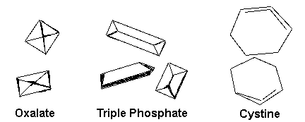
5. Crystals
Common crystals seen even in healthy patients
include calcium oxalate, triple phosphate crystals and amorphous phosphates.

Oxalate crystals in urineTriple phosphate crystals in urineCystine crystals in urine
'임시 보관함' 카테고리의 다른 글
| Test 3. Radiology - US (0) | 2009.01.22 |
|---|---|
| Test 3. Radiology - KUB (0) | 2009.01.22 |
| Test 2. Renal Function Test (0) | 2009.01.22 |
| Test 1. Urinalysis(UA) - Proteinuria. (1) | 2009.01.22 |
| Test 1. Urinalysis(UA) - Hematuria (0) | 2009.01.22 |

